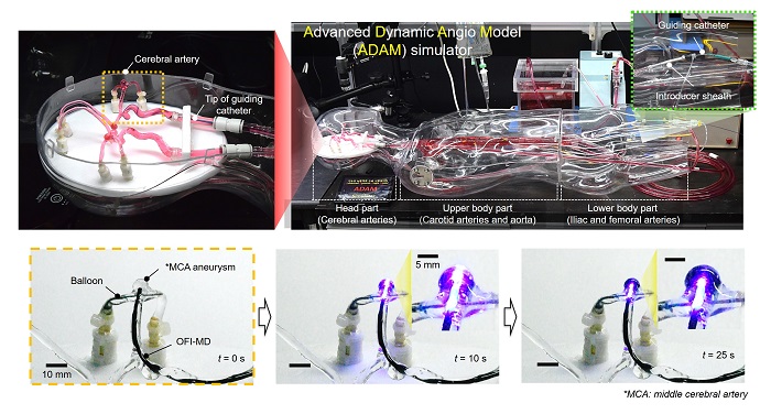
Researchers at Pohang University of Science & Technology (POSTECH) in Korea have developed a new technique to treat cerebral aneurysms. Described in journal Advanced Materials, the treatment involves using a catheter to deliver an alignate hydrogel that is crosslinked in place within the aneurysm using light. The hydrogel is not degradable and can reside within the body for extended periods. As an alternative to coil embolization, the hydrogel stabilizes the aneurysm and reduces the chance of it bursting.
Cerebral aneurysms form when a weak spot in a cerebral artery balloons out, putting pressure on nearby tissues. This ultimately poses a significant risk of a vessel bursting, which frequently proves to be fatal. At present, clinicians may treat such aneurysms through coil embolization, which aims to stabilize the aneurysm with platinum coils. The coils redirect blood flow and reduce pressure within the aneurysm. However, this procedure can be dangerous and can actually cause the vessel to rupture. The coils can also become detached from the vessel.
This new approach involves using an alginate hydrogel to embolize the aneurysm. Such a hydrogel must be liquid when delivered, as it must pass through a narrow catheter and then fill the aneurysm as required, before becoming semi-solid and remaining in place. However, in the tortuous and pulsating environment of a blood vessel, which is full of rushing blood, it can be difficult to control the gel behavior.
These challenges led to the mechanism underlying this latest technology, which incorporates a light-responsive gel which rapidly undergoes a covalent crosslinking reaction when exposed to light. The catheter delivery system incorporates an optical fiber that can deliver light as the gel is extruded, allowing fine control of gel deposition during the procedure. The gel is delivered as microfibers which fill the aneurysm and then undergoes a further ionic crosslinking reaction once in place because of the presence of calcium in the blood.
The gel is not degraded by the body and so should remain in place for a long time. It is also possible to load it with contrast agents so that it can be imaged, both during deposition and then afterwards to monitor whether it is still functioning correctly.
“This research is the first in the world to develop a method that can be used to treat aneurysms by microfibrillating a photocrosslinkable hydrogel microfiber in blood vessels,” said Professor Joonwon Kim, a researcher involved in the study. “It is anticipated these materials will be effectively applicable to many vascular diseases requiring embolization.”


