


A digital microscope is a variation of a traditional optical microscope that uses optics and a digital camera to output an image to a monitor, sometimes by means of software running on a computer. A digital microscope often has its own in-built LED light source, and differs from an optical microscope in that there is no provision to observe the sample directly through an eyepiece. Since the image is focused on the digital circuit, the entire system is designed for the monitor image. The optics for the human eye are omitted.
 Digital microscopes can range from cheap USB digital microscopes to advanced industrial digital microscopes costing tens of thousands of dollars. The low price commercial microscopes normally omit the optics for illumination and are more akin to webcams with a macro lens. For information about stereo microscopes with a digital camera in research and development, see optical microscope.
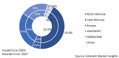
The global digital microscopes market size is estimated to be valued at $1,124.0 million in 2020, and is expected to exhibit a CAGR of 6.5% between 2020 and 2027, according to Coherent Market Insights.
The demand for digital microscopes is expected to increase in the current coronavirus (Covid-19) pandemic. Due to this, pathologists and laboratory worker have started to work from home or remotely. Moreover, digital pathology (digital microscopes) which includes the capturing of high- resolution images of tissue specimen, is one of the factor that has enabled the pathologists to maintain and regulate operations remotely during the pandemic. These digital images can be viewed anywhere and easily shared for second opinions and consults, or in digital teaching sets, overcoming the limitations of working with physical glass slides and microscopes.
MiDiPATHproject
Digital Microscopy in PATHological Anatomy and Cytology) aims to design and develop a set of new digital tools, helping anatomical pathologists refine very serious diseases diagnosis, such as cancers. The tools are specifically designed for the detection of rare elements.
The project brings together the“Centre Hospitalier public du Cotentin” based in Cherbourg, the GREYC laboratories (University of Normandy) based in Caen, and Epinest, a company based in Colombelles. All of the three partners are located in Normandy, France.
This collaborative project is funded by Région Normandie and by the European Union (FEDER).
MiDi PATH project is prize winner of the “ On co – chimie“initiative launched by the Normandie region in partnership with the POLEPHARMA cluster.
Marine Malbert, Responsible RH & Communication at Epinest tell us in detail the purpose of MiDiPATH project in an email interview.
What is the main aim of this project?
The main aim of our project is to make it possible for everyone to have access to early detection of certain types of cancer and to be treated at a very early stage of the disease. This makes it possible to avoid, in certain cases, having to resort to heavy treatments, essential when the detection is done later.
How does this project aim to design and develop a set of new digital tools?
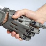
Combining Internet of Things (IOT), AI (artificial intelligence) and machine learning, the use of this combination in the Midipath project will give rise to other products aimed at covering more pathologies.
Tell us in detail about Digital Microscopy and what role does it play in this project?
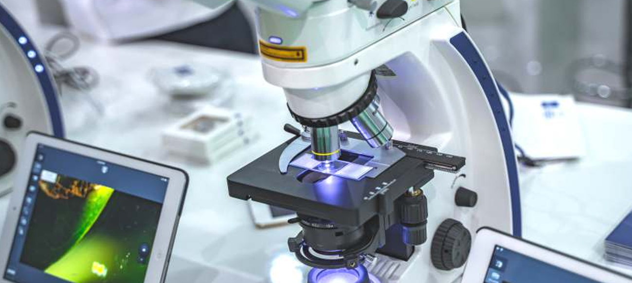
Digital microscopy requires the equipment of classic light microscopy with computerized imaging system. An image of the sample is observed in real-time on a computer screen. This technique includes optical imaging, save feedback information about the microscope status and camera set points. Our project uses digital microscopy in order to receive extremely reliable analyzes, with a much smaller margin of error and unmatched speed.
How does digital microscopy help in diagnosing diseases?
 Digital microscopy allows imaging and measurement at micrometric scale (length, angle, surface, depth, roughness, etc.) of all sample types. Also, looking through an eyepiece for hours is very tiring. Instead of that, the pathologist can sit in a comfortable upright position while viewing a sample on the monitor screen. This makes the working environment easier and more reliable in the analysis
How is it known to be a powerful tool in medicine and bio medical research?
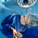
For example, when a surgeon is operating, he sometimes has to take a sample from the patient and needs a quick diagnosis from the pathologist to go further. In some networks of hospitals, the pathologist is not always available, nor on the right site. He is therefore called in emergency, and sometimes spends a considerable amount of time in the car for a few minutes of observation under the microscope. With this technology, the surgeon takes the sample and gives it to a technician who can scan it. The pathologist receives the sample in his mailbox and can complete the protocol to send it back to the surgeon, who can continue the operation. Moreover, several cancer research centers welcome many researchers for two or three years. Each takes hundreds of samples for their studies, which end up in drawers, without any traceability of these slides. To stop such a loss, they will systematically digitize their samples, and will need software like ours to track their activities and feed their biobank. This biobank could be used for future research, in particular by reducing their cost, and by limiting the need for the production of new slides.
 Developments in tech and engineering have allowed the creation of alternatives to high cost diagnostic devices? Your say in this?
A very important factor to remember in this field is the considerable technological advance in recent years in the computing capabilities of personal machines. With advances in algorithms and neural networks we can reach with our technology the same results or even better avoiding big investment in super calculators.
A digital microscope has the same functions as a typical compound microscope. What are its major differences?
Digital microscopes have higher magnification than many optical microscopes because of the use of the computer monitor size to determine the magnification. In addition, digital microscopes provide very high quality images since they project the image directly onto the camera. Other features are also important such as high dynamic range (HDR) for better contrast and color depth, more image texture, speed and of course storage capacity which allows pathologists to reference and examine the images to create detailed reports which gives us the ability to perform deep analysis and identification of rare elements.
Describe the designing of tools for the detection of rare elements?
Producing detection tools means training AI algorithms for all possible scenarios which is deep learning. The bulk of the development is this Neural Network ‘training’. This process allows the tool to display a virtual slide and the keystone is that the robot can understand exactly what is asked. The algorithm retains corrections, refines its search, gains in precision, and at some point, the model is robust. The classification that it automatically suggests to the expert is then at least as precise as a manual classification, and above all, exhaustive, since it takes into account all the pixels of the image. The algorithm can be switched to routine use, and the time savings become considerable. The amount of information sent to the doctor is drastically reduced. The information is concentrated, made more reliable, makes the diagnosis faster and is put at the service of the doctor. Because there is no question of supplanting his diagnosis, nor his expertise.
Is your digital microscopy mobile- based?
Midipath is a set of analysis tools that uses digital microscopy technology. Midipath itself is cross-platform and therefore mobile-based. This means that if the system such as the digital microscope is mobile, then the solution is mobile too.
Can a digital microscopy replace a traditional microscopy? If so, why? Your opinion on this?
It’s a revolution that the world of medicine i s going through. Innovations make it possible to digitize, store, classify and share these slides with a quality of resolution and increasing analysis, which equals or exceeds the precision of the microscope. Storage spaces and internet connections are now secured and sophisticated enough to meet tools such as Midipath. Our opinion is that the mechanics that are put in place have been already evolved. Through the vector of education alone , the next generation of pathologists will work primarily on digitized environment.