


Heart tissue can be imaged in real-time during keyhole procedures using a new optical ultrasound needle developed by researchers at Queen Mary University of London (QMUL) and UCL. The revolutionary technology has been successfully used for minimally invasive heart surgery in pigs, giving an unprecedented, high-resolution view of soft tissues up to 2.5 cm in front of the instrument, inside the body.
Doctors currently rely on external ultrasound probes combined with pre-operative imaging scans to visualise soft tissue and organs during keyhole procedures as the miniature surgical instruments used do not support internal ultrasound imaging.
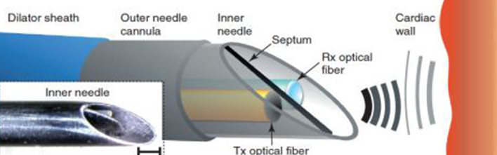
For the study, published today in Light: Science & Applications, the team of surgeons, engineers, physicists and material chemists designed and built the optical ultrasound technology to fit into existing single-use medical devices, such as a needle.
“The optical ultrasound needle is perfect for procedures where there is a small tissue target that is hard to see during keyhole surgery using current methods and missing it could have disastrous consequences,” said Dr Malcolm Finlay, study co-lead and consultant cardiologist at QMUL and Barts Heart Centre.
The team developed the all-optical ultrasound imaging technology for use in a clinical setting over four years. They made sure it was sensitive enough to image centimetre-scale depths of tissues when moving; it fitted into the existing clinical workflow and worked inside the body.
“This is the first demonstration of all-optical ultrasound imaging in a clinically realistic environment. Using inexpensive optical fibres, we have been able to achieve high resolution imaging using needle tips under 1 mm. We now hope to replicate this success across a number of other clinical applications where minimally invasive surgical techniques are being used,” explained study co-lead, Dr Adrien Desjardins (Wellcome EPSRC Centre for Interventional and Surgical Sciences at UCL).
The technology uses a miniature optical fibre encased within a customised clinical needle to deliver a brief pulse of light which generates ultrasonic pulses. Reflections of these ultrasonic pulses from tissue are detected by a sensor on a second optical fibre, giving real-time ultrasound imaging to guide surgery.
“The whole process happens extremely quickly, giving an unprecedented real-time view of soft tissue. It provides doctors with a live image with a resolution of 64 microns, which is the equivalent of only nine red blood cells, and its fantastic sensitivity allows us to readily differentiate soft tissues,” said study co-author, Dr Richard Colchester (UCL Medical Physics & Biomedical Engineering).