


With a wide range of products, Agfa Healthcare constantly aims to improve image quality and workflow using fractional multi-scale image processing, explains the latest white paper
Fractional Multiscale Processing (FMP) is the new mathematical substructure of Agfa HealthCare’s image processing software, which further decomposes image components into elementary fractions for separate processing. FMP results in a more accurate multi-scale enhancement model, a balanced participation of all filter kernel pixels in the enhancement process, and better preservation of low-contrast details next to high-contrast steps.
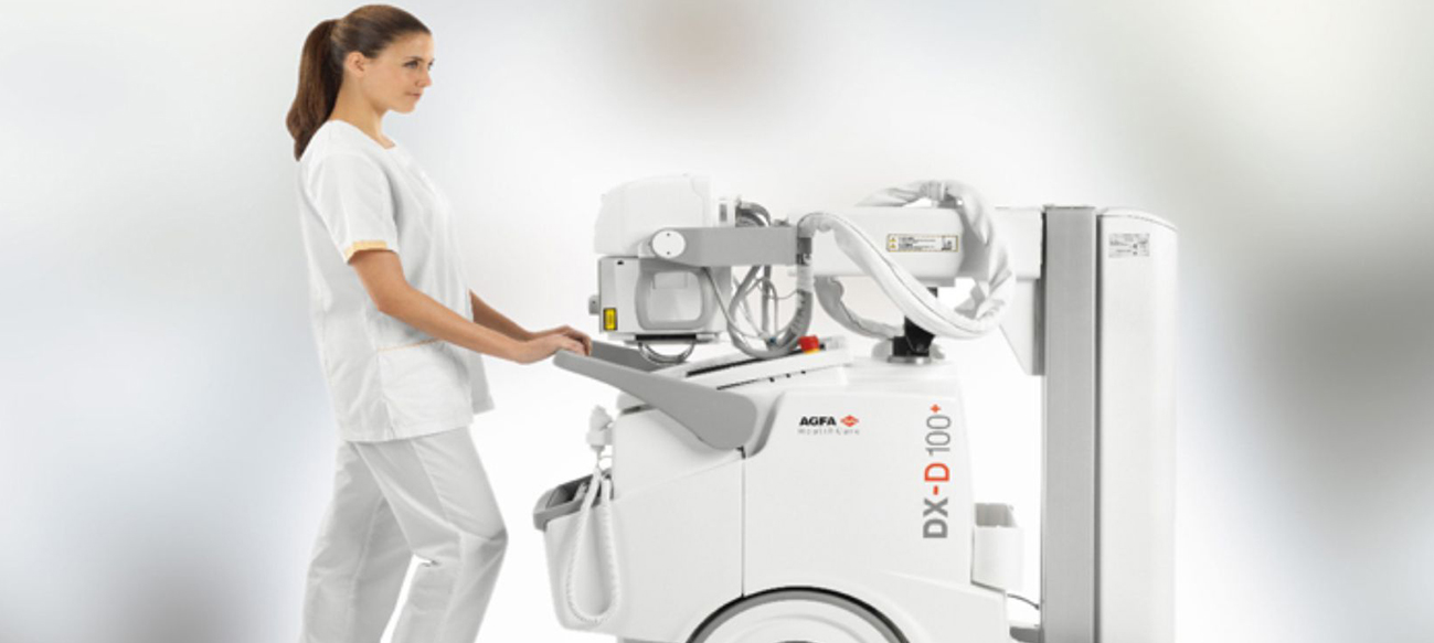
With the advent of mobile digital radiography (DR) systems, combines with an ageing population, there has been a continual increase in the percentage of bedside (portable) check radiographs carried out in hospitals. In some cases, up to 50 per cent of in-hospital digital radiography procedures are now bedside chest exams.
Anti-scatter grids are normally recommended for use with chest radiography in order to improve image quality. Using grids can result in improved contrast detail by reducing the amount of scatter radiation reaching the detector. This is particularly true for medium- to large-sized patients.
But, delivering acceptable image quality from bedside imaging can often be challenging for a technologist, due to equipment and exposure limitations as well as patient pathology .
Agfa HealthCare’s MUSICA image processing is designed to optimize detail contrast under all conditions, whether or not a grid is used. Thus, there is no need for it to mimic the effect of a grid. In this regard, it is conceptually different from conventional image processing, and can process chest images taken with or without a grid, without distinction, using the same algorithm.
 In its latest white paper, Agfa demonstrates MUSICA’s advanced Fractional Multiscale Processing (FMP) technology for these (difficult) bedside chest exposures, and shows how Agfa HealthCare uses state-of-the-art technology to improve the delivery of quality daily care for critically ill patients, as stated by a participating radiologist.
Grid Limitations:
Although using grids is optimal from a physics standpoint, in a bedside imaging setting there are a number of resulting challenges:
Grids need to be properly centered and positioned
- Grids get damaged over time
- Grids may create aliasing artefacts in the images
- Grids typically require a higher radiation dose
- Grids require longer exposure times
Because of these time- and effort-consuming requirements, the use of anti-scatter grids is often avoided in bedside chest radiography.
Chest Radiography
Agfa HealthCare was the first manufacturer to develop image processing algorithms specifically for chest radiography, and for many years MUSICA has optimized non-grid bedside image contrast.
While other technologies attempt to produce an image that resembles the equivalent image taken with a physical anti-scatter grid, MUSICA intrinsically performs scatter subtraction based on an analysis of the image frequencies. Detail contrast can thus be improved, almost up to the level of a properly exposed grid image.
Introduced with MUSICA3, Fractional Multiscale Processing (FMP) further optimizes lung vessel detail, while minimizing the effects of noise and scatter radiation.
In addition to the Fractional Multiscale Processing, MUSICA allows specific tuning or parameter adjustment for non-grid chest imaging, providing additional enhancement. These parameters are versatile and, unlike other products on the market, no system calibration or other pre-requisite is required. The MUSICA concept thus supports greater flexibility to adapt to customer-specific preferences.
The MUSICA3 Chest+ package includes an extra parameter set for optimal bedside chest image quality and facilitates a mix of grid and non-grid imaging workflows.
However, it is to be noted that this application does not replace grid exams in all circumstances (nor does any other technology). Exams taken with a grid under optimal conditions (non-bedside images) can still be superior.
Clinical Image Quality Study
An internal bench test was performed using chest phantoms to simulate both normal-weight and obese patients. This study showed that MUSICA3 chest processing with parameter enhancement could potentially improve the image quality of non-grid chest images to nearly that of a standard MUSICA (Genrad) processed grid image (with a dose reduction factor of 1 .6 for the non-grid images).
However, this phantom testing did not reveal how pathology would be affected by the extra enhancement, and thus what degree of enhancement would be acceptable for radiologists in actual clinical use, depending on patient size and variable X-ray doses. Therefore, a study based on clinical images, including readings by radiologists, was initiated. An elaborated study design was used to derive the optimal MUSICA3 parameter settings, as well as contextual information.
For this purpose, clinical, non-grid bedside chest images from various ICU units were collected from a total of five different hospital sites in the USA, Germany and Belgium. A sample of 25 patient cases was used for evaluation, including DR and CR technology, as well as a representative dose range, different patient sizes and various pathologies.
‘For processing’ raw images from these patient cases were reprocessed with established processing (MUSICA2 Genrad) and with the newest version of Agfa HealthCare’s multi-scale processing (MUSICA3 Chest, including FMP) at default settings, with three different levels of processing enhancement (weak-moderate-strong). The assessment was carried out by six experienced radiologists (two each from the USA, Germany and Belgium).
Improvement in images
MUSICA3 multi-scale processing yields significant improvement in image contract for bedside chest images of normal-weight to obese patients taken without anti-scatter grid. This is particularly noticeable with obese patients. Grid artefacts, long exposure times and un-diagnostic images due to misaligned grids can be avoided.
For non-grid bedside chest images, a statistically significant improvement was seen when using fractional multi-scale processing (MUSICA3) at the minimum and moderate enhancement settings over standard multi-scale image processing (MUSICA2) across all readers and patient sizes (thin to obese).
An even greater improvement was seen when the thin patients were excluded. For normal-weight to obese patients, using minimum enhancement, 81 per cent of the images were rated better than the standard multi-scale processing.
With the moderate enhancement, 77 per cent of the images were rated better. The improvement was more pronounced for images done at higher kVp as well as for images done with lower doses. Image enhancement can be easily optimized using the MUSICA image processing adjustment parameters.
Also, regarding the MUSICA3 default processing for chest, the moderate enhancement was shown to offer a statistically significant improvement for non-grid bedside chest images. Commercially, this MUSICA3 chest enhancement image processing is referred to as ‘Chest+’ .
MUSICA3 Chest+ vs. anti-scatter grids
As 25 non-grid bedside chest images were evaluated comparing MUSICA3 Chest+ to MUSICA2 Genrad image processing, these images were further compared to images from the same patient acquired on another day using a physical anti-scatter grid (ratio 6:1) and increased X-ray dose, and processed using MUSICA 2 Genrad image processing.
While the technologists were trained on the proper use of an anti-scatter grid, this comparison points to many of the typical shortcomings of chest exams taken under these conditions, including grid handling and alignment, variation in patient position and dose (with grid and without grid), and the time difference (and possible pathology change) between the two exposures.
Readings of these image sets were carried out by two experienced radiologists from the hospitals from which the bedside chest images were acquired (specifically, from the ICUs of two typical, larger German hospitals). Both DR and CR images were included. Features for daily control in ICU were identified by the readers, and the images were rated on a quality scale from 1 to 10 (absolute scoring). The overall image quality difference between MUSICA3 Chest+ and MUSICA2 Genrad processing was proven to be significant by means of a T-test with a confidence level of 95 per cent.
Clinical Practice: Conclusion
For bedside chest images, the clinical experience confirmed that MUSICA3 Chest+ was clearly preferred over MUSICA2 Genrad image processing; the MUSICA3 Chest+ non-grid images were rated significantly higher than the MUSICA2 Genrad images carried out with or without a grid.
Specifically, the radiologists felt that the improvement in lung field detail achieved with the MUSICA3 Chest+ image processing without a grid was greater than that achieved using MUSICA2 Genrad processing, even if a grid was used and at an increased dose.
The fact that using a grid and approximately 1.6 times higher dose yields little or no improvement in image quality for the bedside examinations may be attributed to the practical circumstances (and the shortcomings) of grid handling in this situation. In practice, the results for images taken of a patient in an X-ray room with a wall stand, more controlled conditions and different (higher) doses could be expected to yield more favorable results for the grid images.
However, for mobile non-grid imaging, optimal lung field visualization as provided by MUSICA3 Chest+ was seen by the radiologists as the most important aspect of a bedside chest examination. This potentially outweighs the additional improvement of the mediastinum gained by using a physical anti-scatter grid with MUSICA2 Genrad processing at a higher (1 .6x) exposure.